Home
/ Leg Muscle Diagram Side : Ontogenetic Scaling Patterns And Functional Anatomy Of The Pelvic Limb Musculature In Emus Dromaius Novaehollandiae Peerj - The thigh (proximal lower limb) muscles are arranged into three compartments :
Leg Muscle Diagram Side : Ontogenetic Scaling Patterns And Functional Anatomy Of The Pelvic Limb Musculature In Emus Dromaius Novaehollandiae Peerj - The thigh (proximal lower limb) muscles are arranged into three compartments :
Leg Muscle Diagram Side : Ontogenetic Scaling Patterns And Functional Anatomy Of The Pelvic Limb Musculature In Emus Dromaius Novaehollandiae Peerj - The thigh (proximal lower limb) muscles are arranged into three compartments :. Related posts of lower leg muscles diagram muscle anatomy neck. They are attached to the femur (thighbone), tibia (shinbone), and fibula (calf bone) by fibrous tissues called ligaments. It is located toward the middle of the lower leg. The tibia, also known as the shin bone, is the stronger and larger of the two. Observe the leg muscle diagram posted above and notice that there are many parts in the muscles.
This chart is beautifully illustrated and offers the most comprehensive look at the muscles of the human leg available. Medial compartment, also known as adductor compartment; The lower leg is made up of two very strong, long bone—the tibia and the fibula. Start with the gesture then focus on drawing the forms of the quads. Leg muscle pain from medications.
Muscles Of The Lower Limb Calf Muscle Anatomy Leg Muscles Anatomy Body Muscle Anatomy from i.pinimg.com These muscles are found on the front and back sides of the lower leg. Vector illustration informative medical scheme. The accompanying muscle diagram further reveals the positions of the muscles in this pose. The fibularis longus originates from the head and upper lateral surface of the fibula, runs in a bony groove along the bottom of the foot to attach on the other side at the base of the first metatarsal and the neighboring medial cunieform bone, and acts to evert the. Because the leg has many different muscles, it is vulnerable to several different types of muscle strains. This muscle is located on the side of the leg and is actually on the other side of the large toe which it is responsible for moving. Statins and leg pain are a well known combination. Some of the more common ones are:
The lower leg is made up of two very strong, long bone—the tibia and the fibula.
Many medications can cause muscle cramps. As you rotate away from your front leg keep your gaze on the hand that is in the air. The lower leg is made up of two very strong, long bone—the tibia and the fibula. The medialis is the muscle that the sartorius wraps behind before it comes back around to the side of the tibia. The accompanying muscle diagram further reveals the positions of the muscles in this pose. These pictures of this page are about:names of leg muscles diagram. In the leg, muscle strains happen when a muscle is either stretched beyond its limits or forced into extreme contraction. This image shows the muscles of our body and displays them on both male and female diagram showing: Your leg muscles are some of the hardest working muscles in your body. The muscles that make up the quadriceps are the strongest and leanest of all muscles in the body. The leg muscles diagram, will point out if the issue is with any tissue or with the bone. Below the rectus femoris and largely hidden by it is the vastus intermedius. This site contains information about leg muscle diagram side view.
Place your hand on your front leg or floor as you sit back into your front hip with a straight back. But if the pain started after changing a medication or a dose, it is worthwhile looking into. The fibularis longus originates from the head and upper lateral surface of the fibula, runs in a bony groove along the bottom of the foot to attach on the other side at the base of the first metatarsal and the neighboring medial cunieform bone, and acts to evert the. Leg muscle pain from medications. Medial compartment, also known as adductor compartment;
Muscles Of The Knee Anatomy Pictures And Information from www.innerbody.com In the leg, muscle strains happen when a muscle is either stretched beyond its limits or forced into extreme contraction. Most leg pain results from wear and tear, overuse, or injuries in joints or bones or in muscles, ligaments, tendons or other soft tissues. The adductors work virtually any time your legs are active, whether for standing, squatting, lunging, and most other leg moves. Some nerves of the sacral plexus innervate this area, namely the superficial. Detailed anterior, lateral and posterior views.men sports fitness training. Leg muscle anatomical structure, labeled front, side, and back view diagrams. Some common causes of leg pain include: Detailed anterior, lateral and posterior views.men sports fitness training.
Leg muscles diagram muscle diagram.
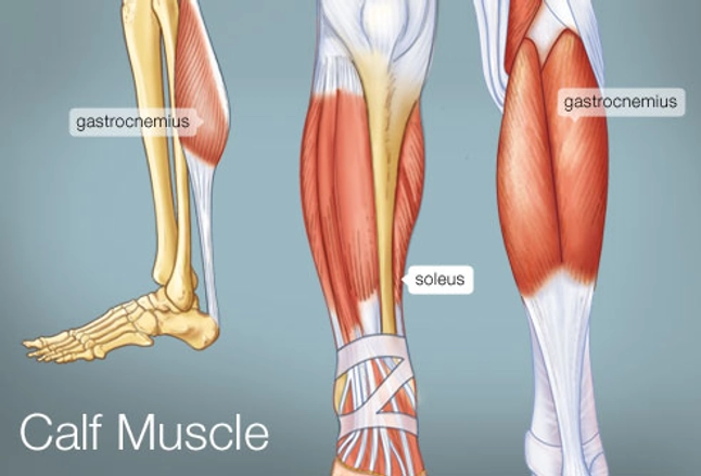
Some types of leg pain can be traced to problems in your lower spine. A muscle strain is a stretch or tear of muscle fibers. The muscles of the leg anatomy chart shows in every possible view the way that the muscles and other pieces of the leg work together in motion and flexibility. Pain in your calf or thigh can be caused by muscle cramps, a pulled or strained muscle, or issues related to your nerves. Place your hand on your front leg or floor as you sit back into your front hip with a straight back. Muscle anatomy neck 12 photos of the muscle anatomy neck dog neck muscle anatomy, front neck muscle anatomy, muscle anatomy neck, muscle anatomy of neck and shoulder, neck muscle anatomy chart, human muscles, dog neck muscle anatomy, front neck muscle anatomy, muscle anatomy neck, muscle anatomy of neck and shoulder, neck. This chart is beautifully illustrated and offers the most comprehensive look at the muscles of the human leg available. Anterior compartment, also known as the extensor compartment; Related posts of muscles and tendons of the leg muscle anatomy diagram. The long head arises from a common tendon with semitendinosus from the superior medial quadrant of the posterior portion of the ischial tuberosity. The short head originates from the lateral lip of linea aspera and. A muscle located on the back portion of the lower leg, being one of the two major muscles that make up the calf:the flexing of this muscle during walking and bending of the knee creates traction on the femur, pulling it toward the tibia in the lower leg and causing the knee to bend. It is located toward the middle of the lower leg.
Some of the more common ones are: The largest muscle masses in the leg are present in the thigh and the calf. A muscle along the outside of the leg that bends the foot out at the ankle. The leg muscles diagram, will point out if the issue is with any tissue or with the bone. The popliteus works on the knee while the other three are associated with the foot and ankle.
The Calf Muscle Human Anatomy Diagram Function Location from img.webmd.com Some types of leg pain can be traced to problems in your lower spine. Most leg pain results from wear and tear, overuse, or injuries in joints or bones or in muscles, ligaments, tendons or other soft tissues. Leg pain can also be caused by blood clots, varicose veins or poor circulation. Related posts of muscles and tendons of the leg muscle anatomy diagram. The fibula, or calf bone, is smaller and is located on the outside of the lower leg. They are attached to the femur (thighbone), tibia (shinbone), and fibula (calf bone) by fibrous tissues called ligaments. Human muscle system, the muscles of the human body that work the skeletal system, that are under voluntary control, and that are concerned with movement, posture, and balance. The fibularis longus originates from the head and upper lateral surface of the fibula, runs in a bony groove along the bottom of the foot to attach on the other side at the base of the first metatarsal and the neighboring medial cunieform bone, and acts to evert the.
A muscle strain is a stretch or tear of muscle fibers.
The following diagram illustrates the actions of the terms adduction, abduction, flexion and extension at the different joints. Included are more than a dozen illustrations like the vastus. The thigh (proximal lower limb) muscles are arranged into three compartments : Your leg muscles are some of the hardest working muscles in your body. Human muscle system, the muscles of the human body that work the skeletal system, that are under voluntary control, and that are concerned with movement, posture, and balance. The medialis is the muscle that the sartorius wraps behind before it comes back around to the side of the tibia. Because the leg has many different muscles, it is vulnerable to several different types of muscle strains. The largest muscle masses in the leg are present in the thigh and the calf. This chart is beautifully illustrated and offers the most comprehensive look at the muscles of the human leg available. The main muscle in this area of the leg is the gastrocnemius, which gives the calf a bulging muscular appearance. Observe the leg muscle diagram posted above and notice that there are many parts in the muscles. The fibula, or calf bone, is smaller and is located on the outside of the lower leg. But if the pain started after changing a medication or a dose, it is worthwhile looking into.
The muscles in the front allow for leg muscle diagram. These muscles are found on the front and back sides of the lower leg.


 muscles are arranged into three compartments :)